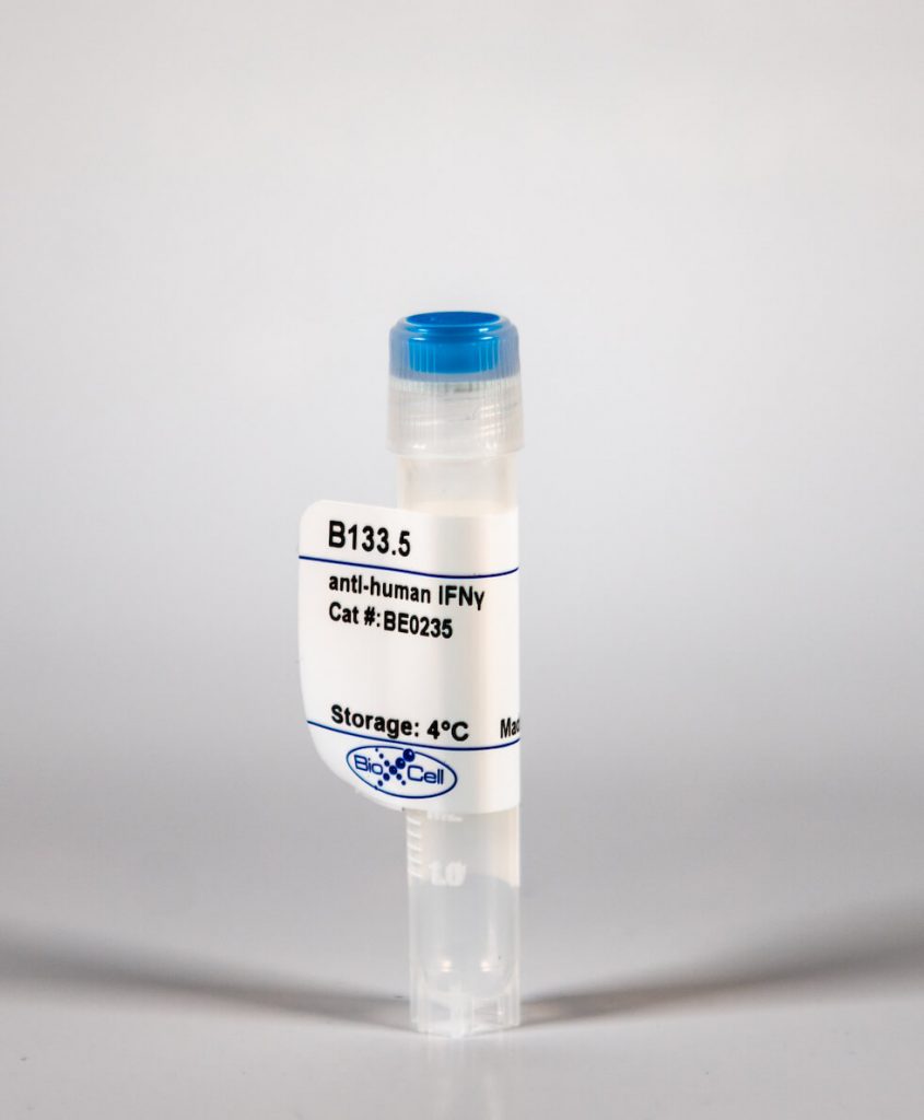
InVivoMab anti-human IFNγ
| Clone | B133.5 | ||||||||||||
|---|---|---|---|---|---|---|---|---|---|---|---|---|---|
| Catalog # | BE0235 | ||||||||||||
| Category | InVivoMab Antibodies | ||||||||||||
| Price |
|
The B133.5 monoclonal antibody reacts with human IFNγ (interferon gamma) a 20 kDa soluble pleiotropic cytokine and the sole member of the type II class of interferons. IFNγ is primarily produced by activated lymphocytes including T, B, NK cells, and ILCs. IFNγ exerts immunoregulatory, anti-proliferative, anti-viral, and proinflammatory activities and plays an important role in activation, growth, and differentiation of T and B lymphocytes, macrophages, NK cells and other non-hematopoietic cell types. Additionally, IFNγ induces the production of cytokines, Fc receptor, and adhesion molecules and up-regulates MHC class I and II antigen expression by antigen presenting cells during an immune response. IFNγ has also been shown to modulate macrophage effector functions, influence isotype switching and induce the secretion of immunoglobulins by B cells. IFNγ signals through the IFN gamma receptor which exists as a heterodimer composed of CD119 (IFN gamma receptor 1) and AF-1 (IFN gamma receptor 2). The IFNγ receptor is expressed ubiquitously on almost all cell types with the exception of mature erythrocytes. The B133.5 antibody is a neutralizing antibody.'
| Isotype | Mouse IgG1 |
| Recommended Isotype Control(s) | InVivoMAb mouse IgG1 isotype control, unknown specificity(BE0083) |
| Recommended InVivoPure Dilution Buffer | InVivoPure pH 7.0 Dilution Buffer(IP0070) |
| Immunogen | Recombinant human IFNγ |
| Reported Applications | in vitro IFNγ neutralization |
| Endotoxin |
|
| Purity |
|
| Formulation |
|
| Sterility | 0.2 μM filtered |
| Production | Purified from tissue culture supernatant in an animal free facility |
| Purification | Protein A |
| Storage | The antibody solution should be stored at the stock concentration at 4°C. Do not freeze. |
| RRID | AB_2687717 |
| Molecular Weight | 150 kDa |
InVivoMAb anti-human IFNγ (Clone: B133.5)
Mitson-Salazar, A., et al. (2015). "Hematopoietic prostaglandin D synthase defines a proeosinophilic pathogenic effector human T2 cell subpopulation with enhanced function." J Allergy Clin Immunol. pii: S0091-6749(15)01186-0. PubMedBACKGROUND: IL-5+ pathogenic effector TH2 (peTH2) cells are a TH2 cell subpopulation with enhanced proinflammatory function that has largely been characterized in murine models of allergic inflammation. OBJECTIVE: We sought to identify phenotype markers for human peTH2 cells and characterize their function in patients with allergic eosinophilic inflammatory diseases. METHODS: Patients with eosinophilic gastrointestinal disease (EGID), patients with atopic dermatitis (AD), and nonatopic healthy control (NA) subjects were enrolled. peTH2 and conventional TH2 (cTH2) cell phenotype, function, and cytokine production were analyzed by using flow cytometry. Confirmatory gene expression was measured by using quantitative RT-PCR. Prostaglandin D2 levels were measured with ELISA. Gut TH2 cells were obtained by means of esophagogastroduodenoscopy. RESULTS: peTH2 cells were identified as chemoattractant receptor-homologous molecule expressed on TH2 cells-positive (CRTH2+), hematopoietic prostaglandin D synthase-positive CD161hi CD4 T cells. peTH2 cells expressed significantly greater IL-5 and IL-13 than did hematopoietic prostaglandin D synthase-negative and CD161- cTH2 cells. peTH2 cells were highly correlated with blood eosinophilia (r = 0.78-0.98) and were present in 30- to 40-fold greater numbers in subjects with EGID and those with AD versus NA subjects. Relative to cTH2 cells, peTH2 cells preferentially expressed receptors for thymic stromal lymphopoietin, IL-25, and IL-33 and demonstrated greater responsiveness to these innate pro-TH2 cytokines. peTH2 but not cTH2 cells produced prostaglandin D2. In patients with EGID and those with AD, peTH2 cells expressed gut- and skin-homing receptors, respectively. There were significantly greater numbers of peTH2 cells in gut tissue from patients with EGID versus NA subjects. CONCLUSION: peTH2 cells are the primary functional proinflammatory human TH2 cell subpopulation underlying allergic eosinophilic inflammation. The unambiguous phenotypic identification of human peTH2 cells provides a powerful tool to track these cells in future pathogenesis studies and clinical trials.
하단영역
(ZIP code : 07532) 902, 9F Hanwha bizmetro 1-cha, 551-17 YangcheonRo,
Gayang-dong, Gangseo-Gu, Seoul, Korea
TEL : 02-2013-8240
FAX : 02-2013-8243
Email : bioxcell@bioxcell.co.kr
Copyright by 2020 BioXCell Korea. All Rights Reserved.
- BioXCell Korea
- Opening hours
- Monday - Friday 9AM - 6PM
- Follow Us
-


