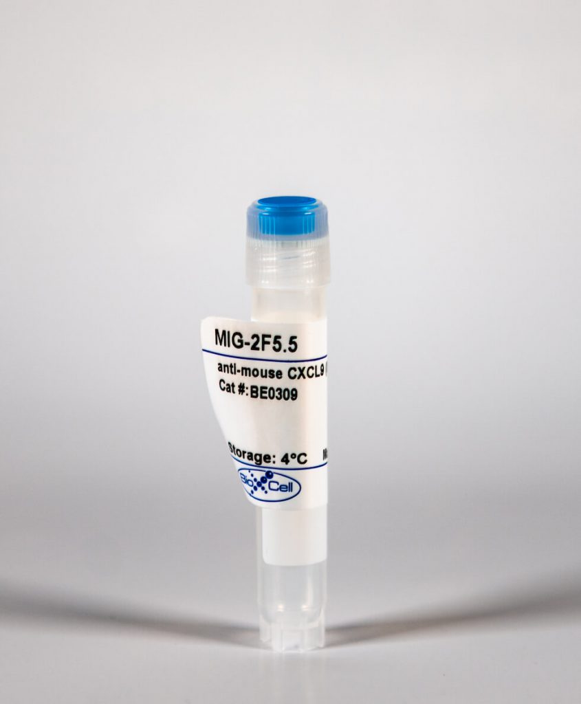
InVivoMAb anti-mouse CXCL9 (MIG)
| Clone | MIG-2F5.5 | ||||||||||||
|---|---|---|---|---|---|---|---|---|---|---|---|---|---|
| Catalog # | BE0309 | ||||||||||||
| Category | InVivoMab Antibodies | ||||||||||||
| Price |
|
The MIG-2F5.5 monoclonal antibody reacts with mouse CXCL9 also known as MIG. CXCL9 is a chemotactic cytokine that belongs to the CXC subfamily of chemokines. CXCL9 is expressed on monocytes, macrophages, hepatocytes, endothelial cells, and primary glial cells in response to IFNγ stimulation. CXCL9 has been shown to be a chemoattractant for resting memory and activated CD4+ and CD8+ T cells, and NK cells expressing its receptor, CXCR3. Binding of CXCL9 to CXCR3 induces various cellular responses, including integrin activation, cytoskeletal changes and chemotactic migration.
| Isotype | Armenian Hamster IgG, κ |
| Recommended Isotype Control(s) | InVivoMAb polyclonal Armenian hamster IgG |
| Recommended Dilution Buffer | InVivoPure™ pH 7.0 Dilution Buffer |
| Reported Applications |
|
| Formulation |
|
| Endotoxin |
|
| Purity |
|
| Sterility | 0.2 μM filtered |
| Production | Purified from tissue culture supernatant in an animal free facility |
| Purification | Protein A |
| RRID | AB_2736989 |
| Molecular Weight | 150 kDa |
| Storage | The antibody solution should be stored at the stock concentration at 4°C. Do not freeze. |
INVIVOMAB ANTI-MOUSE CXCL9 (MIG) (CLONE: MIG-2F5.5)
Xiong, Y., et al. (2016). “T-bet Regulates Natural Regulatory T Cell Afferent Lymphatic Migration and Suppressive Function.” J Immunol 196(6): 2526-2540. PubMed
T-bet is essential for natural regulatory T cells (nTreg) to regulate Th1 inflammation, but whether T-bet controls other Treg functions after entering the inflammatory site is unknown. In an islet allograft model, T-bet(-/-) nTreg, but not induced Treg, failed to prolong graft survival as effectively as wild-type Treg. T-bet(-/-) nTreg had no functional deficiency in vitro but failed to home from the graft to draining lymph nodes (dLN) as efficiently as wild type. T-bet regulated expression of adhesion- and migration-related molecules, influencing nTreg distribution in tissues, so that T-bet(-/-) nTreg remained in the grafts rather than migrating to lymphatics and dLN. In contrast, both wild-type and T-bet(-/-) CD4(+) conventional T cells and induced Treg migrated normally toward afferent lymphatics. T-bet(-/-) nTreg displayed instability in the graft, failing to suppress Ag-specific CD4(+) T cells and prevent their infiltration into the graft and dLN. Thus, T-bet regulates nTreg migration into afferent lymphatics and dLN and consequently their suppressive stability in vivo.
Jin, J. O., et al. (2013). “Innate immune signaling induces interleukin-7 production from salivary gland cells and accelerates the development of primary Sjogren’s syndrome in a mouse model.” PLoS One 8(10): e77605. PubMed
Elevated IL-7 in the target tissues is closely associated with multiple autoimmune disorders, including Sjogren’s syndrome (SS). We recently found that IL-7 plays an essential role in the development and onset of primary SS (pSS) in C57BL/6.NOD-Aec1Aec2 mice, a well-defined mouse model of primary SS. However, environmental signals that cause excessive IL-7 production are not well-characterized. Innate immune signaling plays a critical role in shaping the adaptive immune responses including autoimmune responses. We and others have previously shown that innate immune signaling can induce IL-7 expression in lungs and intestines of C57BL/6 mice. In this study, we characterized the effects of poly I:C, a double-stranded RNA analog and toll-like receptor 3 agonist, on the induction of IL-7 expression in salivary glands and on pSS development. We showed that poly I:C administration to C57BL/6 mice rapidly induced IL-7 expression in the salivary glands in a type 1 IFN- and IFN-gamma-dependent manner. Moreover, poly I:C-induced IL-7 contributed to the optimal up-regulation of CXCL9 in the salivary glands, which may subsequently promote recruitment of more IFN-gamma-producing T cells. Repeated administration of poly I:C to C57BL/6.NOD-Aec1Aec2 mice accelerated the development of SS-like exocrinopathy, and this effect was abolished by the blockade of IL-7 receptor signaling with a neutralizing antibody. Finally, poly I:C or a combination of IFN-alpha and IFN-gamma induced IL-7 gene expression and protein production in a human salivary gland epithelial cell line. Hence, we demonstrate that IL-7 expression in the salivary gland cells can be induced by poly I:C and delineate a crucial mechanism by which innate immune signals facilitate the development of pSS, which is through induction of IL-7 in the target tissues.
Sung, J. H., et al. (2012). “Chemokine guidance of central memory T cells is critical for antiviral recall responses in lymph nodes.” Cell 150(6): 1249-1263. PubMed
A defining feature of vertebrate immunity is the acquisition of immunological memory, which confers enhanced protection against pathogens by mechanisms that are incompletely understood. Here, we compared responses by virus-specific naive T cells (T(N)) and central memory T cells (T(CM)) to viral antigen challenge in lymph nodes (LNs). In steady-state LNs, both T cell subsets localized in the deep T cell area and interacted similarly with antigen-presenting dendritic cells. However, upon entry of lymph-borne virus, only T(CM) relocalized rapidly and efficiently toward the outermost LN regions in the medullary, interfollicular, and subcapsular areas where viral infection was initially confined. This rapid peripheralization was coordinated by a cascade of cytokines and chemokines, particularly ligands for T(CM)-expressed CXCR3. Consequently, in vivo recall responses to viral infection by CXCR3-deficient T(CM) were markedly compromised, indicating that early antigen detection afforded by intranodal chemokine guidance of T(CM) is essential for efficient antiviral memory.
하단영역
(ZIP code : 07532) 902, 9F Hanwha bizmetro 1-cha, 551-17 YangcheonRo,
Gayang-dong, Gangseo-Gu, Seoul, Korea
TEL : 02-2013-8240
FAX : 02-2013-8243
Email : bioxcell@bioxcell.co.kr
Copyright by 2020 BioXCell Korea. All Rights Reserved.
- BioXCell Korea
- Opening hours
- Monday - Friday 9AM - 6PM
- Follow Us
-


