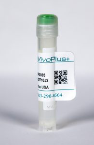
InVivoPlus mouse IgG2a isotype control, unknown specificity
| Clone | C1.18.4 | ||||||||||
|---|---|---|---|---|---|---|---|---|---|---|---|
| Catalog # | BP0085 | ||||||||||
| Category | Isotype Controls | ||||||||||
| Price |
|
The C1.18.4 monoclonal antibody is ideal for use as a non-reactive isotype-matched control for mouse IgG2a antibodies in most in vivo and in vitro applications.
| Isotype | Mouse IgG2a, κ |
| Recommended Dilution Buffer | InVivoPure™ pH 7.0 Dilution Buffer |
| Formulation |
|
| Endotoxin |
|
| Aggregation |
|
| Purity |
|
| Sterility | 0.2 μM filtered |
| Production | Purified from tissue culture supernatant in an animal free facility |
| Purification | Protein G |
| RRID | AB_1107771 |
| Molecular Weight | 150 kDa |
| Murine Pathogen Test Results |
|
| Storage | The antibody solution should be stored at the stock concentration at 4°C. Do not freeze. |
INVIVOPLUS MOUSE IGG2A ISOTYPE CONTROL; UNKNOWN SPECIFICITY (CLONE: C1.18.4)
Carmi, Y., et al. (2015). “Allogeneic IgG combined with dendritic cell stimuli induce antitumour T-cell immunity.” Nature 521(7550): 99-104. PubMed
Whereas cancers grow within host tissues and evade host immunity through immune-editing and immunosuppression, tumours are rarely transmissible between individuals. Much like transplanted allogeneic organs, allogeneic tumours are reliably rejected by host T cells, even when the tumour and host share the same major histocompatibility complex alleles, the most potent determinants of transplant rejection. How such tumour-eradicating immunity is initiated remains unknown, although elucidating this process could provide the basis for inducing similar responses against naturally arising tumours. Here we find that allogeneic tumour rejection is initiated in mice by naturally occurring tumour-binding IgG antibodies, which enable dendritic cells (DCs) to internalize tumour antigens and subsequently activate tumour-reactive T cells. We exploited this mechanism to treat autologous and autochthonous tumours successfully. Either systemic administration of DCs loaded with allogeneic-IgG-coated tumour cells or intratumoral injection of allogeneic IgG in combination with DC stimuli induced potent T-cell-mediated antitumour immune responses, resulting in tumour eradication in mouse models of melanoma, pancreas, lung and breast cancer. Moreover, this strategy led to eradication of distant tumours and metastases, as well as the injected primary tumours. To assess the clinical relevance of these findings, we studied antibodies and cells from patients with lung cancer. T cells from these patients responded vigorously to autologous tumour antigens after culture with allogeneic-IgG-loaded DCs, recapitulating our findings in mice. These results reveal that tumour-binding allogeneic IgG can induce powerful antitumour immunity that can be exploited for cancer immunotherapy.
Nakatsukasa, H., et al. (2015). “The DNA-binding inhibitor Id3 regulates IL-9 production in CD4(+) T cells.” Nat Immunol 16(10): 1077-1084. PubMed
The molecular mechanisms by which signaling via transforming growth factor-beta (TGF-beta) and interleukin 4 (IL-4) control the differentiation of CD4(+) IL-9-producing helper T cells (TH9 cells) remain incompletely understood. We found here that the DNA-binding inhibitor Id3 regulated TH9 differentiation, as deletion of Id3 increased IL-9 production from CD4(+) T cells. Mechanistically, TGF-beta1 and IL-4 downregulated Id3 expression, and this process required the kinase TAK1. A reduction in Id3 expression enhanced binding of the transcription factors E2A and GATA-3 to the Il9 promoter region, which promoted Il9 transcription. Notably, Id3-mediated control of TH9 differentiation regulated anti-tumor immunity in an experimental melanoma-bearing model in vivo and also in human CD4(+) T cells in vitro. Thus, our study reveals a previously unrecognized TAK1-Id3-E2A-GATA-3 pathway that regulates TH9 differentiation.
Bulliard, Y., et al. (2013). “Activating Fc gamma receptors contribute to the antitumor activities of immunoregulatory receptor-targeting antibodies.” J Exp Med 210(9): 1685-1693. PubMed
Fc gamma receptor (FcgammaR) coengagement can facilitate antibody-mediated receptor activation in target cells. In particular, agonistic antibodies that target tumor necrosis factor receptor (TNFR) family members have shown dependence on expression of the inhibitory FcgammaR, FcgammaRIIB. It remains unclear if engagement of FcgammaRIIB also extends to the activities of antibodies targeting immunoregulatory TNFRs expressed by T cells. We have explored the requirement for activating and inhibitory FcgammaRs for the antitumor effects of antibodies targeting the TNFR glucocorticoid-induced TNFR-related protein (GITR; TNFRSF18; CD357) expressed on activated and regulatory T cells (T reg cells). We found that although FcgammaRIIB was dispensable for the in vivo efficacy of anti-GITR antibodies, in contrast, activating FcgammaRs were essential. Surprisingly, the dependence on activating FcgammaRs extended to an antibody targeting the non-TNFR receptor CTLA-4 (CD152) that acts as a negative regulator of T cell immunity. We define a common mechanism that correlated with tumor efficacy, whereby antibodies that coengaged activating FcgammaRs expressed by tumor-associated leukocytes facilitated the selective elimination of intratumoral T cell populations, particularly T reg cells. These findings may have broad implications for antibody engineering efforts aimed at enhancing the therapeutic activity of immunomodulatory antibodies.
Kerzerho, J., et al. (2013). “Programmed cell death ligand 2 regulates TH9 differentiation and induction of chronic airway hyperreactivity.” J Allergy Clin Immunol 131(4): 1048-1057, 1057 e1041-1042. PubMed
BACKGROUND: Asthma is defined as a chronic inflammatory disease of the airways; however, the underlying physiologic and immunologic processes are not fully understood. OBJECTIVE: The aim of this study was to determine whether TH9 cells develop in vivo in a model of chronic airway hyperreactivity (AHR) and what factors control this development. METHOD: We have developed a novel chronic allergen exposure model using the clinically relevant antigen Aspergillus fumigatus to determine the time kinetics of TH9 development in vivo. RESULTS: TH9 cells were detectable in the lungs after chronic allergen exposure. The number of TH9 cells directly correlated with the severity of AHR, and anti-IL-9 treatment decreased airway inflammation. Moreover, we have identified programmed cell death ligand (PD-L) 2 as a negative regulator of TH9 cell differentiation. Lack of PD-L2 was associated with significantly increased TGF-beta and IL-1alpha levels in the lungs, enhanced pulmonary TH9 differentiation, and higher morbidity in the sensitized mice. CONCLUSION: Our findings suggest that PD-L2 plays a pivotal role in the regulation of TH9 cell development in chronic AHR, providing novel strategies for modulating adaptive immunity during chronic allergic responses.
Licona-Limon, P., et al. (2013). “Th9 Cells Drive Host Immunity against Gastrointestinal Worm Infection.” Immunity 39(4): 744-757. PubMed
Type 2 inflammatory cytokines, including interleukin-4 (IL-4), IL-5, IL-9, and IL-13, drive the characteristic features of immunity against parasitic worms and allergens. Whether IL-9 serves an essential role in the initiation of host-protective responses is controversial, and the importance of IL-9- versus IL-4-producing CD4(+) effector T cells in type 2 immunity is incompletely defined. Herein, we generated IL-9-deficient and IL-9-fluorescent reporter mice that demonstrated an essential role for this cytokine in the early type 2 immunity against Nippostrongylus brasiliensis. Whereas T helper 9 (Th9) cells and type 2 innate lymphoid cells (ILC2s) were major sources of infection-induced IL-9 production, the adoptive transfer of Th9 cells, but not Th2 cells, caused rapid worm expulsion, marked basophilia, and increased mast cell numbers in Rag2-deficient hosts. Taken together, our data show a critical and nonredundant role for Th9 cells and IL-9 in host-protective type 2 immunity against parasitic worm infection.
Rayamajhi, M., et al. (2012). “Lung B cells promote early pathogen dissemination and hasten death from inhalation anthrax.” Mucosal Immunol 5(4): 444-454. PubMed
Sampling of mucosal antigens regulates immune responses but may also promote dissemination of mucosal pathogens. Lung dendritic cells (LDCs) capture antigens and traffic them to lung-draining lymph nodes (LDLNs) dependent on the chemokine receptor CCR7 (chemokine (C-C motif) receptor 7). LDCs also capture lung pathogens such as Bacillus anthracis (BA). However, we show here that the initial traffic of BA spores from lungs to LDLNs is largely independent of LDCs and CCR7, occurring instead in association with B cells. BA spores rapidly bound B cells in lungs and cultured mouse and human B cells. Binding was independent of the B-cell receptor (BCR). B cells instilled in the lungs trafficked to LDLNs and BA spore traffic to LDLNs was impaired by B-cell deficiency. Depletion of B cells also delayed death of mice receiving a lethal BA infection. These results suggest that mucosal B cells traffic BA, and possibly other antigens, from lungs to LDLNs.
Schafer, H., et al. (2012). “Myofibroblast-induced tumorigenicity of pancreatic ductal epithelial cells is L1CAM dependent.” Carcinogenesis 33(1): 84-93. PubMed
Pancreatic ductal adenocarcinoma (PDAC) and chronic pancreatitis, representing one risk factor for PDAC, are characterized by a marked desmoplasia enriched of pancreatic myofibroblasts (PMFs). Thus, PMFs are thought to essentially promote pancreatic tumorigenesis. We recently demonstrated that the adhesion molecule L1CAM is involved in epithelial-mesenchymal transition of PMF-cocultured H6c7 human ductal epithelial cells and that L1CAM is expressed already in ductal structures of chronic pancreatitis with even higher elevation in primary tumors and metastases of PDAC patients. This study aimed at investigating whether PMFs and L1CAM drive malignant transformation of pancreatic ductal epithelial cells by enhancing their tumorigenic potential. Cell culture experiments demonstrated that in the presence of PMFs, H6c7 cells exhibit a profound resistance against death ligand-induced apoptosis. This apoptosis protection was similarly observed in H6c7 cells stably overexpressing L1CAM. Intrapancreatic inoculation of H6c7 cells together with PMFs (H6c7co) resulted in tumor formation in 7/8 and liver metastases in 6/8 severe combined immunodeficiency (SCID) mice, whereas no tumors and metastases were detectable after inoculation of H6c7 cells alone. Likewise, tumor outgrowth and metastases resulted from inoculation of L1CAM-overexpressing H6c7 cells in 5/7 and 3/7 SCID mice, respectively, but not from inoculation of mock-transfected H6c7 cells. Treatment of H6c7co tumor-bearing mice with the L1CAM antibody L1-9.3/2a inhibited tumor formation and liver metastasis in 100 and 50%, respectively, of the treated animals. Overall, these data provide new insights into the mechanisms of how PMFs and L1CAM contribute to malignant transformation of pancreatic ductal epithelial cells in early stages of pancreatic tumorigenesis.
Lamere, M. W., et al. (2011). “Regulation of antinucleoprotein IgG by systemic vaccination and its effect on influenza virus clearance.” J Virol 85(10): 5027-5035. PubMed
Seasonal influenza epidemics recur due to antigenic drift of envelope glycoprotein antigens and immune evasion of circulating viruses. Additionally, antigenic shift can lead to influenza pandemics. Thus, a universal vaccine that protects against multiple influenza virus strains could alleviate the continuing impact of this virus on human health. In mice, accelerated clearance of a new viral strain (cross-protection) can be elicited by prior infection (heterosubtypic immunity) or by immunization with the highly conserved internal nucleoprotein (NP). Both heterosubtypic immunity and NP-immune protection require antibody production. Here, we show that systemic immunization with NP readily accelerated clearance of a 2009 pandemic H1N1 influenza virus isolate in an antibody-dependent manner. However, human immunization with trivalent inactivated influenza virus vaccine (TIV) only rarely and modestly boosted existing levels of anti-NP IgG. Similar results were observed in mice, although the reaction could be enhanced with adjuvants, by adjusting the stoichiometry among NP and other vaccine components, and by increasing the interval between TIV prime and boost. Importantly, mouse heterosubtypic immunity that had waned over several months could be enhanced by injecting purified anti-NP IgG or by boosting with NP protein, correlating with a long-lived increase in anti-NP antibody titers. Thus, current immunization strategies poorly induce NP-immune antibody that is nonetheless capable of contributing to long-lived cross-protection. The high conservation of NP antigen and the known longevity of antibody responses suggest that the antiviral activity of anti-NP IgG may provide a critically needed component of a universal influenza vaccine.
Libbey, J. E., et al. (2011). “Interleukin-6, produced by resident cells of the central nervous system and infiltrating cells, contributes to the development of seizures following viral infection.” J Virol 85(14): 6913-6922. PubMed
Cells that can participate in an innate immune response within the central nervous system (CNS) include infiltrating cells (polymorphonuclear leukocytes , macrophages, and natural killer cells) and resident cells (microglia and sometimes astrocytes). The proinflammatory cytokine interleukin-6 (IL-6) is produced by all of these cells and has been implicated in the development of behavioral seizures in the Theiler’s murine encephalomyelitis virus (TMEV)-induced seizure model. The assessment, via PCR arrays, of the mRNA expression levels of a large number of chemokines (ligands and receptors) in TMEV-infected and mock-infected C57BL/6 mice both with and without seizures did not clearly demonstrate the involvement of PMNs, monocytes/macrophages, or NK cells in the development of seizures, possibly due to overlapping function of the chemokines. Additionally, C57BL/6 mice unable to recruit or depleted of infiltrating PMNs and NK cells had seizure rates comparable to those of controls following TMEV infection, and therefore PMNs and NK cells do not significantly contribute to seizure development. In contrast, C57BL/6 mice treated with minocycline, which affects monocytes/macrophages, microglial cells, and PMNs, had significantly fewer seizures than controls following TMEV infection, indicating monocytes/macrophages and resident microglial cells are important in seizure development. Irradiated bone marrow chimeric mice that were either IL-6-deficient mice reconstituted with wild-type bone marrow cells or wild-type mice reconstituted with IL-6-deficient bone marrow cells developed significantly fewer behavioral seizures following TMEV infection. Therefore, both resident CNS cells and infiltrating cells are necessary for seizure development.


