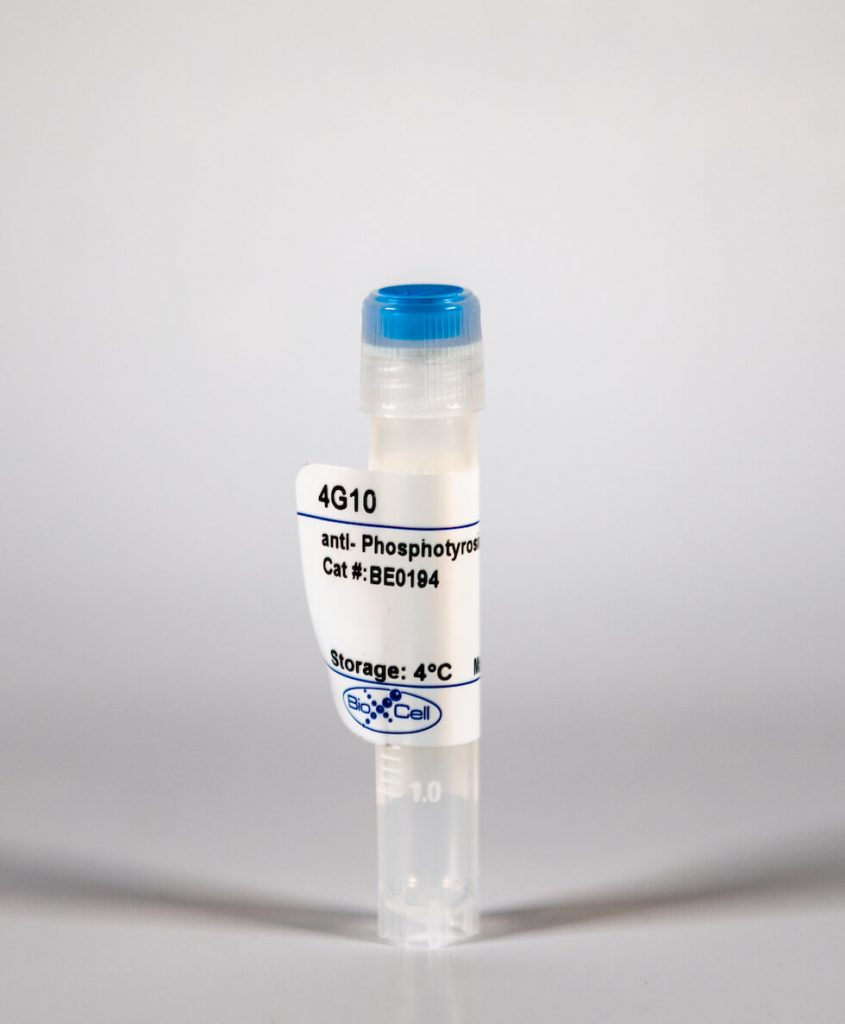
InVivoMab anti-Phosphotyrosine
| Clone | 4G10 | ||||||||||||
|---|---|---|---|---|---|---|---|---|---|---|---|---|---|
| Catalog # | BE0194 | ||||||||||||
| Category | InVivoMab Antibodies | ||||||||||||
| Price |
|
The 4B10 monoclonal antibody reacts with tyrosine phosphorylated proteins in all species.'
| Isotype | Mouse IgG2b |
| Recommended Isotype Control(s) | InVivoMAb mouse IgG2b isotype control, unknown specificity(BE0086) |
| Recommended InVivoPure Dilution Buffer | InVivoPure pH 7.0 Dilution Buffer(IP0070) |
| Immunogen | Phosphotyramine-KLH |
| Reported Applications |
|
| Endotoxin |
|
| Purity |
|
| Formulation |
|
| Sterility | 0.2 μM filtered |
| Production | Purified from tissue culture supernatant in an animal free facility |
| Purification | Protein G |
| Storage | The antibody solution should be stored at the stock concentration at 4°C. Do not freeze. |
| RRID | AB_10949186 |
| Molecular Weight | 150 kDa |
InVivoMAb anti-Phosphotyrosine (Clone: 4G10)
Klammt, C., et al. (2015). "T cell receptor dwell times control the kinase activity of Zap70." Nat Immunol 16(9): 961-969. PubMed
Kinase recruitment to membrane receptors is essential for signal transduction. However, the underlying regulatory mechanisms are poorly understood. We investigated how conformational changes control T cell receptor (TCR) association and activity of the kinase Zap70. Structural analysis showed that TCR binding or phosphorylation of Zap70 triggers a transition from a closed, autoinhibited conformation to an open conformation. Using Zap70 mutants with defined conformations, we found that TCR dwell times controlled Zap70 activity. The closed conformation minimized TCR dwell times and thereby prevented activation by membrane-associated kinases. Parallel recruitment of coreceptor-associated Lck kinase to the TCR ensured Zap70 phosphorylation and stabilized Zap70 TCR binding. Our study suggests that the dynamics of cytosolic enzyme recruitment to the plasma membrane regulate the activity and function of receptors lacking intrinsic catalytic activity.
Whittall, J. J., et al. (2015). "Topology of Streptococcus pneumoniae CpsC, a polysaccharide copolymerase and bacterial protein tyrosine kinase adaptor protein." J Bacteriol 197(1): 120-127. PubMed
In Gram-positive bacteria, tyrosine kinases are split into two proteins, the cytoplasmic tyrosine kinase and a transmembrane adaptor protein. In Streptococcus pneumoniae, this transmembrane adaptor is CpsC, with the C terminus of CpsC critical for interaction and subsequent tyrosine kinase activity of CpsD. Topology predictions suggest that CpsC has two transmembrane domains, with the N and C termini present in the cytoplasm. In order to investigate CpsC topology, we used a chromosomal hemagglutinin (HA)-tagged Cps2C protein in S. pneumoniae strain D39. Incubation of both protoplasts and membranes with carboxypeptidase B (CP-B) resulted in complete degradation of HA-Cps2C in all cases, indicating that the C terminus of Cps2C was likely extracytoplasmic and hence that the protein's topology was not as predicted. Similar results were seen with membranes from S. pneumoniae strain TIGR4, indicating that Cps4C also showed similar topology. A chromosomally encoded fusion of HA-Cps2C and Cps2D was not degraded by CP-B, suggesting that the fusion fixed the C terminus within the cytoplasm. However, capsule synthesis was unaltered by this fusion. Detection of the CpsC C terminus by flow cytometry indicated that it was extracytoplasmic in approximately 30% of cells. Interestingly, a mutant in the protein tyrosine phosphatase CpsB had a significantly greater proportion of positive cells, although this effect was independent of its phosphatase activity. Our data indicate that CpsC possesses a varied topology, with the C terminus flipping across the cytoplasmic membrane, where it interacts with CpsD in order to regulate tyrosine kinase activity.


