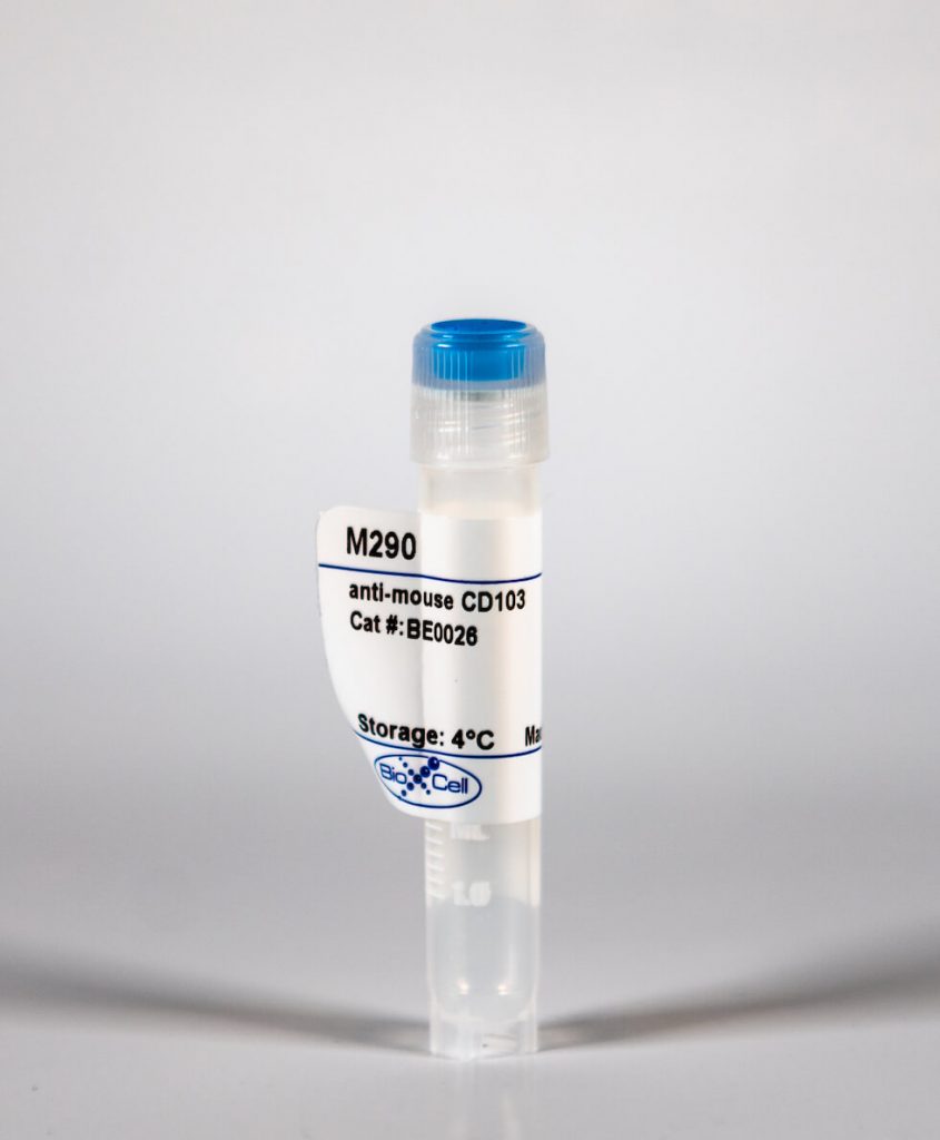
InVivoMab anti-mouse CD103
| Clone | M290 | ||||||||||||
|---|---|---|---|---|---|---|---|---|---|---|---|---|---|
| Catalog # | BE0026 | ||||||||||||
| Category | InVivoMab Antibodies | ||||||||||||
| Price |
|
The M290 monoclonal antibody reacts with mouse CD103 also known as integrin αE (ITGAE). CD103 is an integrin protein that binds integrin beta 7 to form the complete heterodimeric integrin molecule αEβ7. CD103 is expressed widely on intraepithelial lymphocyte (IEL) T cells (both αβ T cells and γδ T cells) and on some peripheral regulatory T cells. It has also been reported on lamina propria T cells. A subset of dendritic cells in the gut mucosa and in mesenteric lymph nodes also expresses CD103. The main ligand for CD103 is E-cadherin, an adhesion molecule expressed by epithelial cells. CD103 is thought to facilitate the interactions of T cells with epithelial cells during T cell maturation and effector functions. The M290 antibody is reported to neutralize CD103 in vivo.
| Isotype | Rat IgG2a, κ |
| Recommended Isotype Control(s) | InVivoMAb rat IgG2a isotype control, anti-trinitrophenol |
| Recommended Dilution Buffer | InVivoPure™ pH 7.0 Dilution Buffer |
| Immunogen | Mouse intestinal epithelial cells |
| Reported Applications |
|
| Formulation |
|
| Endotoxin |
|
| Purity |
|
| Sterility | 0.2 μM filtered |
| Production | Purified from tissue culture supernatant in an animal free facility |
| Purification | Protein G |
| RRID | AB_1107570 |
| Molecular Weight | 150 kDa |
| Storage | The antibody solution should be stored at the stock concentration at 4°C. Do not freeze. |
INVIVOMAB ANTI-MOUSE CD103 (CLONE: M290)
Mang, Y., et al. (2015). “Efficient elimination of CD103-expressing cells by anti-CD103 antibody drug conjugates in immunocompetent mice.” Int Immunopharmacol 24(1): 119-127. PubMed
CD103 plays an important role in the destruction of islet allografts, and previous studies found that a CD103 immunotoxin (M290-Saporin, or M290-SAP) promoted the long-term survival of pancreatic islet allografts. However, systemic toxicity to the host and the bystander effects of M290-SAP obscure the underlying mechanisms of action and restrict its clinical applications. To overcome these shortcomings, anti-CD103 M290 was conjugated to different cytotoxic agents through cleavable or uncleavable linkages to form three distinct antibody-drug conjugates (ADCs): M290-MC-vc-PAB-MMAE, M290-MC-MMAF, and M290-MCC-DM1. The drug-to-antibody ratio (DAR) and the purity of the ADCs were determined by HIC-HPLC and SEC-HPLC, respectively. The binding characteristics, internalization and cytotoxicity of M290 and the corresponding ADCs were evaluated in vitro. The cell depletion efficacies of the various M290-ADCs against CD103-positive cells were then evaluated in vivo. The M290-ADCs maintained the initial binding affinity for the CD103-positive cell surface antigen and then quickly internalized the CD103-positive cell. Surprisingly, all M290-ADCs potently depleted CD103-positive cells in vivo, with high specificity and reduced toxicity. Our findings show that M290-ADCs have potent and selective depletion effects on CD103-expressing cells in immunocompetent mice. These data indicate that M290-ADCs could potentially serve as a therapeutic intervention to block the CD103/E-cadherin pathway.
Mock, J. R., et al. (2014). “Foxp3+ regulatory T cells promote lung epithelial proliferation.” Mucosal Immunol 7(6): 1440-1451. PubMed
Acute respiratory distress syndrome (ARDS) causes significant morbidity and mortality each year. There is a paucity of information regarding the mechanisms necessary for ARDS resolution. Foxp3(+) regulatory T cells (Foxp3(+) T(reg) cells) have been shown to be an important determinant of resolution in an experimental model of lung injury. We demonstrate that intratracheal delivery of endotoxin (lipopolysaccharide) elicits alveolar epithelial damage from which the epithelium undergoes proliferation and repair. Epithelial proliferation coincided with an increase in Foxp3(+) T(reg) cells in the lung during the course of resolution. To dissect the role that Foxp3(+) T(reg) cells exert on epithelial proliferation, we depleted Foxp3(+) T(reg) cells, which led to decreased alveolar epithelial proliferation and delayed lung injury recovery. Furthermore, antibody-mediated blockade of CD103, an integrin, which binds to epithelial expressed E-cadherin decreased Foxp3(+) T(reg) numbers and decreased rates of epithelial proliferation after injury. In a non-inflammatory model of regenerative alveologenesis, left lung pneumonectomy, we found that Foxp3(+) T(reg) cells enhanced epithelial proliferation. Moreover, Foxp3(+) T(reg) cells co-cultured with primary type II alveolar cells (AT2) directly increased AT2 cell proliferation in a CD103-dependent manner. These studies provide evidence of a new and integral role for Foxp3(+) T(reg) cells in repair of the lung epithelium.
Sandoval, F., et al. (2013). “Mucosal imprinting of vaccine-induced CD8(+) T cells is crucial to inhibit the growth of mucosal tumors.” Sci Transl Med 5(172): 172ra120. PubMed
Although many human cancers are located in mucosal sites, most cancer vaccines are tested against subcutaneous tumors in preclinical models. We therefore wondered whether mucosa-specific homing instructions to the immune system might influence mucosal tumor outgrowth. We showed that the growth of orthotopic head and neck or lung cancers was inhibited when a cancer vaccine was delivered by the intranasal mucosal route but not the intramuscular route. This antitumor effect was dependent on CD8(+) T cells. Indeed, only intranasal vaccination elicited mucosal-specific CD8(+) T cells expressing the mucosal integrin CD49a. Blockade of CD49a decreased intratumoral CD8(+) T cell infiltration and the efficacy of cancer vaccine on mucosal tumor. We then showed that after intranasal vaccination, dendritic cells from lung parenchyma, but not those from spleen, induced the expression of CD49a on cocultured specific CD8(+) T cells. Tumor-infiltrating lymphocytes from human mucosal lung cancer also expressed CD49a, which supports the relevance and possible extrapolation of these results in humans. We thus identified a link between the route of vaccination and the induction of a mucosal homing program on induced CD8(+) T cells that controlled their trafficking. Immunization route directly affected the efficacy of the cancer vaccine to control mucosal tumors.


