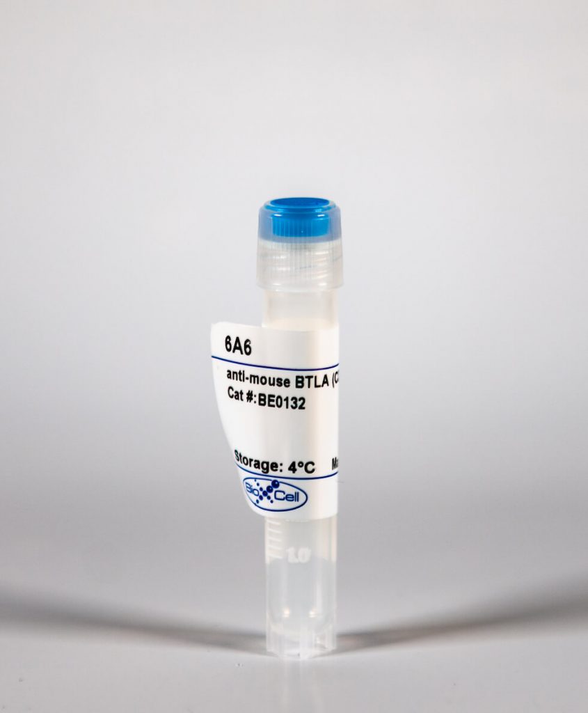
InVivoMAb anti-mouse BTLA (CD272)
| Clone | 6A6 | ||||||||||||
|---|---|---|---|---|---|---|---|---|---|---|---|---|---|
| Catalog # | BE0132 | ||||||||||||
| Category | InVivoMab Antibodies | ||||||||||||
| Price |
|
The 6A6 monoclonal antibody reacts with mouse B- and T-lymphocyte attenuator (BTLA) also known as CD272. BTLA is an Ig superfamily member which is expressed on B cells, T cells, macrophages, dendritic cells, NK cells, and NKT cells. Like PD-1 and CTLA-4, BTLA interacts with a B7 homolog, B7-H4. However, unlike PD-1 and CTLA-4, BTLA displays T cell inhibition via interaction with tumor necrosis family receptors, not just the B7 family of cell surface receptors. BTLA is a ligand for herpes virus entry mediator (HVEM). BTLA-HVEM complexes have been shown to negatively regulate T cell immune responses.
| Isotype | Armenian Hamster IgG, κ |
| Recommended Isotype Control(s) | InVivoMAb polyclonal Armenian hamster IgG |
| Recommended Dilution Buffer | InVivoPure™ pH 7.0 Dilution Buffer |
| Immunogen | C57BL/6 mouse BTLA Ig domain |
| Reported Applications |
|
| Formulation |
|
| Endotoxin |
|
| Purity |
|
| Sterility | 0.2 μM filtered |
| Production | Purified from tissue culture supernatant in an animal free facility |
| Purification | Protein A |
| RRID | AB_10949299 |
| Molecular Weight | 150 kDa |
| Storage | The antibody solution should be stored at the stock concentration at 4°C. Do not freeze. |
InVivoMAb anti-mouse BTLA (CD272) (Clone: 6A6)
Bekiaris, V., et al. (2013). “The inhibitory receptor BTLA controls gammadelta T cell homeostasis and inflammatory responses.” Immunity 39(6): 1082-1094. PubMed
gammadelta T cells rapidly secrete inflammatory cytokines at barrier sites that aid in protection from pathogens, but mechanisms limiting inflammatory damage remain unclear. We found that retinoid-related orphan receptor gamma-t (RORgammat) and interleukin-7 (IL-7) influence gammadelta T cell homeostasis and function by regulating expression of the inhibitory receptor, B and T lymphocyte attenuator (BTLA). The transcription factor RORgammat, via its activating function-2 domain, repressed Btla transcription, whereas IL-7 increased BTLA levels on the cell surface. BTLA expression limited gammadelta T cell numbers and sustained normal gammadelta T cell subset frequencies by restricting IL-7 responsiveness and expansion of the CD27(-)RORgammat(+) population. BTLA also negatively regulated IL-17 and TNF production in CD27(-) gammadelta T cells. Consequently, BTLA-deficient mice exhibit enhanced disease in a gammadelta T cell-dependent model of dermatitis, whereas BTLA agonism reduced inflammation. Therefore, by coordinating expression of BTLA, RORgammat and IL-7 balance suppressive and activation stimuli to regulate gammadelta T cell homeostasis and inflammatory responses.
Nakagomi, D., et al. (2013). “Therapeutic potential of B and T lymphocyte attenuator expressed on CD8+ T cells for contact hypersensitivity.” J Invest Dermatol 133(3): 702-711. PubMed
In the past decade, mechanisms underlying allergic contact dermatitis have been intensively investigated by using contact hypersensitivity (CHS) models in mice. However, the regulatory mechanisms, which could be applicable for the treatment of allergic contact dermatitis, are still largely unknown. To determine the roles of B and T lymphocyte attenuator (BTLA), a CD28 family coinhibitory receptor, in hapten-induced CHS, BTLA-deficient (BTLA(-/-)) mice and littermate wild-type (WT) mice were subjected to DNFB-induced CHS, severe combined immunodeficient (SCID) mice were injected with CD4(+) T cells, and CD8(+) T cells from either WT mice or BTLA(-/-) mice were subjected to CHS. BTLA(-/-) mice showed enhanced DNFB-induced CHS and proliferation and IFN-gamma production of CD8(+) T cells as compared with WT mice. SCID mice injected with WT CD4(+) T cells and BTLA(-/-) CD8(+) T cells exhibited more severe CHS as compared with those injected with WT CD4(+) T cells and WT CD8(+) T cells. On the other hand, SCID mice injected with BTLA(-/-) CD4(+) T cells and WT CD8(+) T cells exhibited similar CHS to those injected with WT CD4(+) T cells and WT CD8(+) T cells. Finally, to evaluate the therapeutic potential of an agonistic agent for BTLA on CHS, the effects of an agonistic anti-BTLA antibody (6A6) on CHS were examined. In vivo injection of 6A6 suppressed DNFB-induced CHS and IFN-gamma production of CD8(+) T cells. Taken together, these results suggest that stimulation of BTLA with agonistic agents has therapeutic potential in CHS.
Steinberg, M. W., et al. (2013). “BTLA interaction with HVEM expressed on CD8(+) T cells promotes survival and memory generation in response to a bacterial infection.” PLoS One 8(10): e77992. PubMed
The B and T lymphocyte attenuator (BTLA) is an Ig super family member that binds to the herpes virus entry mediator (HVEM), a TNF receptor super family (TNFRSF) member. Engagement of BTLA by HVEM triggers inhibitory signals, although recent evidence indicates that BTLA also may act as an activating ligand for HVEM. In this study, we reveal a novel role for the BTLA-HVEM pathway in promoting the survival of activated CD8(+) T cells in the response to an oral microbial infection. Our data show that both BTLA- and HVEM-deficient mice infected with Listeria monocytogenes had significantly reduced numbers of primary effector and memory CD8(+) T cells, despite normal proliferation and expansion compared to controls. In addition, blockade of the BTLA-HVEM interaction early in the response led to significantly reduced numbers of antigen-specific CD8(+) T cells. HVEM expression on the CD8(+) T cells as well as BTLA expression on a cell type other than CD8(+) T lymphocytes, was required. Collectively, our data demonstrate that the function of the BTLA-HVEM pathway is not limited to inhibitory signaling in T lymphocytes, and instead, that BTLA can provide crucial, HVEM-dependent signals that promote survival of antigen activated CD8(+) T cell during bacterial infection.
Tewalt, E. F., et al. (2012). “Lymphatic endothelial cells induce tolerance via PD-L1 and lack of costimulation leading to high-level PD-1 expression on CD8 T cells.” Blood 120(24): 4772-4782. PubMed
Lymphatic endothelial cells (LECs) induce peripheral tolerance by direct presentation to CD8 T cells (T(CD8)). We demonstrate that LECs mediate deletion only via programmed cell death-1 (PD-1) ligand 1, despite expressing ligands for the CD160, B- and T-lymphocyte attenuator, and lymphocyte activation gene-3 inhibitory pathways. LECs induce activation and proliferation of T(CD8), but lack of costimulation through 4-1BB leads to rapid high-level expression of PD-1, which in turn inhibits up-regulation of the high-affinity IL-2 receptor that is necessary for T(CD8) survival. Rescue of tyrosinase-specific T(CD8) by interference with PD-1 or provision of costimulation results in autoimmune vitiligo, demonstrating that LECs are significant, albeit suboptimal, antigen-presenting cells. Because LECs express numerous peripheral tissue antigens, lack of costimulation coupled to rapid high-level up-regulation of inhibitory receptors may be generally important in systemic peripheral tolerance.
Lepenies, B., et al. (2007). “Ligation of B and T lymphocyte attenuator prevents the genesis of experimental cerebral malaria.” J Immunol 179(6): 4093-4100. PubMed
B and T lymphocyte attenuator (BTLA; CD272) is a coinhibitory receptor that is predominantly expressed on T and B cells and dampens T cell activation. In this study, we analyzed the function of BTLA during infection with Plasmodium berghei ANKA. Infection of C57BL/6 mice with this strain leads to sequestration of leukocytes in brain capillaries that is associated with a pathology resembling cerebral malaria in humans. During the course of infection, we found an induction of BTLA in several organs, which was either due to up-regulation of BTLA expression on T cells in the spleen or due to infiltration of BTLA-expressing T cells into the brain. In the brain, we observed a marked induction of BTLA and its ligand herpesvirus entry mediator during cerebral malaria, which was accompanied by an accumulation of predominantly CD8+ T cells, but also CD4+ T cells. Application of an agonistic anti-BTLA mAb caused a significantly reduced incidence of cerebral malaria compared with control mice. Treatment with this Ab also led to a decreased number of T cells that were sequestered in the brain of P. berghei ANKA-infected mice. Our findings indicate that BTLA-herpesvirus entry mediator interactions are functionally involved in T cell regulation during P. berghei ANKA infection of mice and that BTLA is a potential target for therapeutic interventions in severe malaria.


